

Myomectomy
In addition, with a correct interpretation of the images obtained by this ultrasound technology, polycystic ovaries, simple and complex ovarian cysts of different sizes can be diagnosed, the presence of uterine fibroids in their various locations using the endovaginal area to obtain the location of the fibroids. or polyps, if any, and to be able to define the surgical technique that will be used.
Myomas

Uterus with 3 myomas

Extirpated myomas
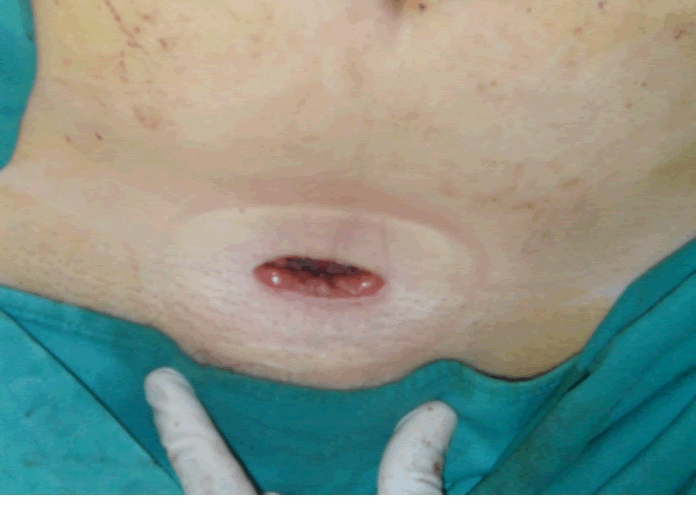
Actual size of the wound
